In Situ Synchrotron X-ray Characterization on the Nucleation and Growth Kinetics of Colloidal Nanopa

A 6-minute read
In this review, the kinetics of the key processes leading to the formation of colloidal nanoparticles are covered, along with the potential of in situ synchrotron X-ray techniques for analyzing the relevant kinetics. In situ high energy synchrotron X-ray techniques that have recently has been developed to provide a fantastic opportunity to quantitatively track the growth trajectories of colloidal nanoparticles in real time under reaction-specific conditions. To elucidate the mechanism of nanoparticle production and establish the related intrinsic kinetic parameters, only of the lost data

Figure 1. Schematic illustration of time-dependent evolution of concentrations of precursor reactant (red), nanoparticle precursor (green), and size of nanoparticles (blue) involved in a typical synthesis of collagen nanoparticles. The major steps are (I) nucleation, (II) early growth, and (III) late growth stages. The background sketches the evolution of NPs from reactive to. The lower part highlights the appropriate in situ X-ray techniques for different evolution periods ( S. Wu et al., 2019 ).
When a synthesis reaction begins, the process of converting the reactant(s) into the NP precursor, which is required for the generation of solid nuclei, will occur. As the process proceeds, more NP precursor is built up until it reaches a supersaturation point , at which case it spontaneously condenses to form solid nuclei in the liquid solution (ie, nucleation stage I in Figure 1). In a liquid solution of supersaturated NP precursor, a solid nucleus is regarded as the smallest stable solid particle local. To ascertain chemical environment of target elements, in situ X-ray absorption fine structure (XAFS) can be used to examine the transformation of reactant into NP precursor (pre-nucleation). The emergence of solid nuclei is better characterized by the size-sensitive in situ SAXS (ie, condensation of NP precursor, or nucleation). The additional reaction promotes NP growth, or the enlargement of nuclei to NPs. Due to the high surface energy of microscopic NPs, Ostwald ripening and NP coalescence may take place at the early development stage II (Figure 1) together with growth. In order to follow the growth kinetics, in situ SAXS is the most suitable method. The development of NPs and intra-particle reconstruction take control at the late development stage III (Figure 1) of the process. The associated growth kinetics and reconstruction evolution can be observed using both wide-angle X-ray scattering (WAXS) and in situ synchrotron SAXS. In order to follow the growth kinetics, in situ SAXS is the most suitable method. The development of NPs and intra-particle reconstruction take control at the late development stage III (Figure 1) of the process.The associated growth kinetics and reconstruction evolution can be observed using both wide-angle X-ray scattering (WAXS) and in situ synchrotron SAXS. In order to follow the growth kinetics, in situ SAXS is the most suitable method. The development of NPs and intra-particle reconstruction take control at the late development stage III (Figure 1) of the process. The associated growth kinetics and reconstruction evolution can be observed using both wide-angle X-ray scattering (WAXS) and in situ synchrotron SAXS.The associated growth kinetics and reconstruction evolution can be observed using both wide-angle X-ray scattering (WAXS) and in situ synchrotron SAXS. Growth kinetics and reconstruction evolution can be observed using both wide-angle X-ray scattering (WAXS) and in situ synchrotron SAXS.
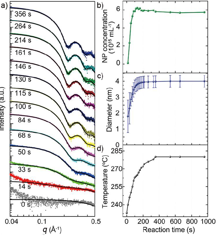
Figure 2. a) In situ SAXS patterns of growing Pd CNPs recorded at various times. The dotted plots and solid plots are experimental data and the corresponding fitting results, respectively. The extracted time-dependent b) concentration and c) size (symbols) and size distribution (error bars) of the Pd CNPs determined from analysis of the in situ SAXS patterns. d) Time dependent temperature of the synthesis solution (L. Wu et al., 2018) .
In Figure 2a, the time-dependent SAXS patterns for a typical synthesis reaction using only TOP surfactant (Pd:TOP molar ratio = 1:5) are shown. The SAXS patterns were examined to determine the NPs' size, concentration, and size distribution (number of NPs in 1 mL solution) (Figures 2b and 2c). Fast nucleation occurred in a period of 50 seconds when the temperature was raised to 230 °C, and thereafter there was continuous growth of NPs from 2.8 to 3.7 nm in diameter (Figure 2d). At the growth stage, slow nucleation continued to take place, with a nucleation rate of 2.4 x 10 13 mL -1 s- 1 up to 100 s, which was significantly slower than the rate of the initial fast nucleation ( 1.22 x 10 14 mL -1 s -1 ). Due to Ostwald ripening, which caused the NPs to concentrate in size, the number of NPs gradually dropped at around 200s.

References
[1] Wu, S., Li, M., & Sun, Y. (2019). In Situ Synchrotron X-ray Characterization Shining Light on the Nucleation and Growth Kinetics of Colloidal Nanoparticles. Angewandte Chemie - International Edition , 58 ( 27 ), 8987–8995. https://doi.org/10.1002/anie.201900690
[2] Wu, L., Lian, H., Willis, JJ, Goodman, ED, McKay, IS, Qin, J., Tassone, CJ, & Cargnello, M. (2018). Tuning Precursor Reactivity toward Nanometer-Size Control in Palladium Nanoparticles Studied by in Situ Small Angle X-ray Scattering. Chemistry of Materials , 30 (3), 1127–1135. https://doi.org/10.1021/acs.chemmater.7b05186